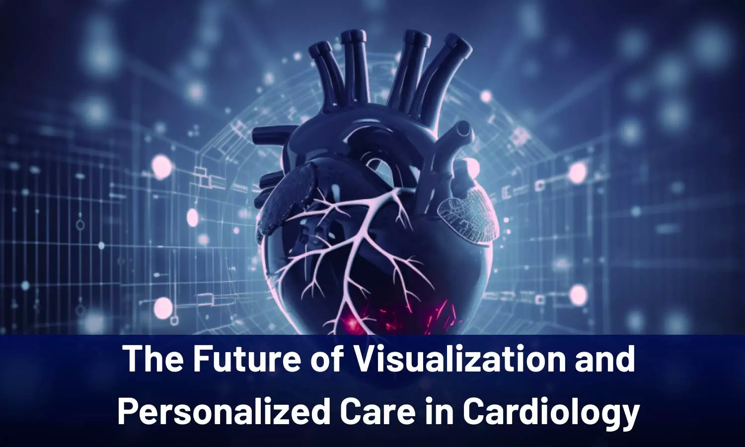

A
recent special article reviewed future
applications of visual representations and concepts through artificial
intelligence (AI) aimed at enhancing cardiovascular care, medical education,
and cardiac research.
This
special article is published in December 2025 in the American Heart Journal.
This news article
elaborates on the role of technological innovations, including artificial
intelligence, virtual reality, and 3D modeling, in transforming future visual
art applications for personalized cardiac care.
The
evolution of futuristic visual arts is driven by the integration of AI and
machine learning (ML) alongside immersive experiences that incorporate virtual
and augmented realities. These technological integrations create endless
possibilities for personalized medicine, medical education, and research.
Artificial Intelligence (AI) and
Machine Learning (ML) Staged to Revolutionize Personalized Treatment
Decision-Making
Over
the last decade, statistical, computational, and physical 3D modeling
methodologies have evolved to support clinical decision-making, surgical
planning, and overall pathophysiological understanding. These models are
constructed by processing routinely acquired clinical 3D imaging data, such as
CT, MR, and echocardiography. The evolution of AI tools in cardiovascular care
has created opportunities for improved diagnostics, individualized treatment,
and outcome prediction through ML applications. Cardiologists can utilize
text-to-image AI-generative art platforms to create more engaging and
personalized educational content on specific cardiac pathologies and
procedures, adjusting the visuals to the patient’s literacy level and
sociocultural preferences.
Immersive Technology for Procedural
Planning
Immersive
technologies such as Augmented Reality (AR) and Virtual Reality (VR) are set to
revolutionize procedural planning. VR environments could potentially enable
cardiologists and cardiac surgeons to interact with a patient’s cardiovascular
anatomy before a procedure, thereby decreasing procedural complications. HeartFlow FFRCT, the first FDA-cleared
simulation platform for cardiovascular modeling, uses patient-specific anatomy
derived from CT images and non-personalized parameters to objectively measure
coronary blood flow. Additionally, Extended Reality (ER) imaging models have
been utilized in catheterization laboratories to create real-time 3D digital
holograms from rotational angiography, echocardiography and electroanatomic
mapping for preoperative planning.
Visualizing Cardiac Electrical
Activity and Scar Tissue
In
electrophysiology, AI-generative art can be used to generate artistic
representations of the heart’s electrical activity from real-time cardiac 3D
electroanatomic mapping and ECGs. These customized, patient-derived interactive
arts enable cardiologists to visualize arrhythmogenic patterns and scar tissue
derived from computational modeling, decreasing the reliance on traditional
fluoroscopic methods. Furthermore, visual representation techniques allow
research into non-invasive fractional flow reserve measurements using CT
angiography data, reducing the need for invasive procedures.
The Role of 3D Printing in Surgical
Navigation
Advanced
visualization techniques, particularly 3D printing, demonstrate potential to
revolutionize healthcare. The first clinical intracardiac application involved
creating mitral valve biomodels
using 3D echocardiographic datasets. This model will likely be expanded to
structural heart disease and aortic aneurysms, which assist in surgical
navigation.
Integrating Visual Art in Future
Education & Public Health Outreach
The
integration of arts into medical education is a growing trend, with many
institutions incorporating visual arts to enhance visual perception skills,
develop empathy, and cultivate humanistic aspects of practice. For patients, VR
platforms could be beneficial in helping them visualize their coronary artery
calcium, thereby ultimately improving adherence to necessary lifestyle
modifications. Public health advocates can utilize visual arts, such as public
exhibits and AI-generative content, to engage communities in heart attack
awareness and the prevention of cardiovascular diseases.
Complementing
Visual Art with Technology in Cardiology– The Future Seems Bright
The integration of AI and immersive
visual technologies will be instrumental in providing personalized healthcare. Giving patients immersive
experiences—such as explaining a heart transplant or the placement of a
WATCHMAN device through a physical, interactive environment—will simplify
complex procedures and offer comfort and understanding. These advancements are
expected to become commonplace within a few years, demonstrating the influence
of visual art and technology on improved surgical techniques, tailored care,
and the enhancement of both the scientific precision and the humanistic aspects
of cardiovascular medicine.
Reference: Ugoala O, Ebubechukwu U, Mares AC, Okeke C, Anosike U,
Tamirisa KP, Obuobi S, Gibson CM. Visual art and representation in cardiology:
Past, present, and future. Am Heart J. 2025 Dec;290:201-215. doi:
10.1016/j.ahj.2025.06.016. Epub 2025 Jun 27. PMID: 40582478.
For regular cardiology updates from recent journals, kindly follow our WhatsApp group