Persistent inflammation of the endometrial mucosa is known
as chronic endometritis (CE). This condition is characterized by the
microscopic identification of plasma cells in the endometrial stroma. Numerous
bacteria, primarily gram-negative and intracellular (such as Enterococcus
faecalis, Mycoplasma, ureaplasma Chlamydia, Escherichia coli, and Streptococcus
spp.), have been associated with the development of CE, but some cases of
abacterial CE are described.
Although frequently asymptomatic, women with CE often
complain of vaginal discharge, dyspareunia, pelvic pain, and abnormal uterine
bleeding. Furthermore, multiple studies have shown women with primary
infertility, recurrent implantation failure (RIF), and recurrent pregnancy loss
(RPL) to have a higher prevalence of CE compared to the general population, suggesting
a potential correlation between CE and reproductive disorders. The deleterious
impact of CE on fertility is often attributed to the aberrant infiltration of
plasma cells with the consequent release of antibodies and cytokines, but it is
still subject to debate. Notably, women with CE also display altered
endometrial expression of genes encoding for proteins implicated in the
inflammatory response, proliferation, and apoptosis.
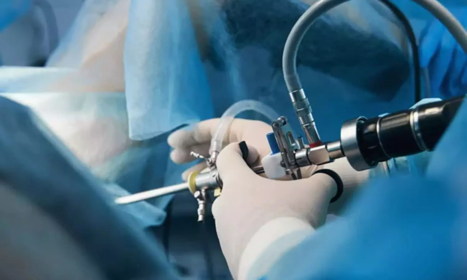
Hysteroscopy is the current gold standard technique for both
the diagnosis and treatment of intracavitary and endocervical lesions.
Hysteroscopy has already been shown to have high diagnostic accuracy in women
with endometrial polyps, submucosal fibroids, hyperplasia, and endometrial
cancer. As a result, hysteroscopy is considered as an effective first-line
diagnostic technique for women with infertility, in whom the presence of
endometrial pathology may negatively influence the endometrial receptivity for
the embryo. In this respect, a comprehensive examination of the endometrial
cavity, including targeted biopsies if needed, is enabled through a
hysteroscopic approach.
To assess the diagnostic accuracy of current hysteroscopic
criteria compared with histopathological analysis (with or without additional
immunohistochemistry) for the detection of chronic endometritis MEDLINE,
Scopus, SciELO, Embase, ClinicalTrials.gov, Cochrane Central Register of
Controlled Trials, LILACS, conference proceedings, and international controlled
trials registries were searched without date limit or language restrictions.
Studies were selected if they were randomized, prospective,
or retrospective and estimated the diagnostic accuracy of hysteroscopy for
chronic endometritis by comparing hysteroscopic criteria with histopathological
(with or without immunohistochemistry) diagnosis. Primary outcomes were the
diagnostic odds ratio, area under the summary receiver operating characteristic
curve, sensitivity, and specificity. Positive and negative likelihood ratios
were secondary outcomes.
Diagnostic accuracy meta-analysis was conducted following
the Preferred Reporting Items for Systematic Reviews and MetaAnalyses and
Synthesizing Evidence from Diagnostic Accuracy Tests recommendations and
Synthesizing Evidence from Diagnostic Accuracy Tests methodological guidelines.
Quality assessment was conducted using the Quality Assessment Tool for
Diagnostic Accuracy Studies. Publication bias was evaluated with Deeks funnel
plot asymmetry test.
Thirteen studies compared available hysteroscopic criteria
(stromal edema, diffuse or focal hyperemia, “strawberry aspect,”
micropolyposis) with subsequent histopathological analysis of endometrial
sampling. After pooling all the studies, the diagnostic odds ratio was 40 (95%
confidence interval, 12-133). The evaluated area under summary receiver
operating characteristic curve was 0.93 (95% confidence interval, 0.90-0.95),
correlating with very high diagnostic accuracy. Sensitivity and specificity
were 84% (95% confidence interval, 0.68-0.93) and 89% (95% confidence interval,
0.75-0.95), respectively. In addition, the positive and negative likelihood
ratios were 7.4 (95% confidence interval 3.2-17.0) and 0.19 (95% confidence
interval, 0.09-0.39), respectively.
This systematic review and DTA metaanalysis shows that the
use of currently available hysteroscopic features for diagnosing CE has high
accuracy. Data could be computed from all 13 papers that were part of the
systematic review. Hysteroscopy has results comparable to the gold standard of
histopathology, as seen by the area under the SROC curve, which indicates high
accuracy for the index test. A high PLR (>5.0) and low NLR (<0.2) are
additional requirements for a diagnostic test to be deemed effective. The PLR
score of 8.3 in this meta-analysis indicates that women meeting at least one
hysteroscopic criterion are nearly 9 times more likely to test positive for
endometritis at histopathology. Moreover, in women without hysteroscopic
suggestive findings, the NLR value of 0.20 represents a 5-fold reduction in the
likelihood of having CE. CIs for the evaluated outcomes overlapped, suggesting
good quality evidence.
This systematic review and DTA metaanalysis on both
infertile and noninfertile women show that the current hysteroscopic criteria
for diagnosing CE demonstrate accuracy prior to histopathological confirmation.
Accordingly, the absence of hysteroscopic findings suggestive of the presence
of CE would not need histologic confirmation and would make supplemental biopsy
of more limited yield. However, the limitations of this study and reviewed
evidence do not allow to draw strict conclusions. In fact, when clinical
suspicion is high, hysteroscopic results are unclear, or patient anxiety or
history warrants additional confirmation, histopathologic confirmation is
recommended. Hysteroscopic biopsy and/or second look confirmation may also be
extremely important for assessing the therapeutic response to antibiotic
regimens and for identifying CE instances that have resistant features and
necessitate additional histopathological assessment. Moreover, integrating
RTPCR could increase diagnostic precision, especially in uncertain cases.
Additional studies are required to propose the integration of molecular
diagnostics as a complementary standard and to clarify the role of hysteroscopic
targeted endometrial biopsy, relative to blind techniques, in obtaining optimal
samples for subsequent histopathological analysis in patients with suspected
CE.
Source: Gaetano Riemma, John Preston Parry, Pasquale De
Franciscis; American Journal of Obstetrics & Gynecology JULY 2025
https://doi.org/10.1016/j.ajog.2025.03.005